
Beyond Bone Density: Don’t Wait Until You Break

How Echolight REMS and the Beyond Your Scan Experience Are Creating a More Proactive Path to Bone Health
By Margaret Wallis-Duffy, RMT, CPCA Founder, Wallis for Wellness and the Preventative Health Awareness Movement
For most people, bone health is invisible until it is not.
There is no warning light on the dashboard. Bone loss usually does not hurt. You can feel well, stay active and move through life with no idea that your skeletal strength may be changing beneath the surface.
Then comes the fracture.
That is the moment our healthcare system often begins paying close attention. But after 34 years as a Registered Massage Therapist with a background in Kinesiology, working in musculoskeletal and preventative health, I believe we need to ask a better question:
Why are we waiting until people break before helping them understand what is holding them up?
That question is at the heart of Wallis for Wellness: Beyond Your Scan.
The Beyond Your Scan Experience combines Health Canada–licensed Echolight REMS technology with something technology alone can never provide: clear education, practical resources, informed self-advocacy and connection to an integrative healthcare team.
Because receiving a result is not the finish line. It is the starting point.
The opportunity gap in bone health
Most people reach peak bone mass by their late twenties or early thirties. After that, the goal shifts: preserve what you have, reduce avoidable losses and continue supporting the muscles, balance and mobility that protect you from falls and fractures.
Yet under the current Canadian clinical guideline, baseline bone mineral density testing with dual-energy X-ray absorptiometry, commonly called DXA or DEXA, begins at age 70 for adults without risk factors. Testing is recommended earlier for people whose age, fracture history, medications or medical conditions place them at greater risk.
Osteoporosis Canada’s 2023 guideline recommends testing at 65 when one risk factor is present, and between 50 and 64 following a previous fracture or when two or more risk factors are present.
Those recommendations matter. DXA remains the established clinical reference standard and an essential part of osteoporosis care.
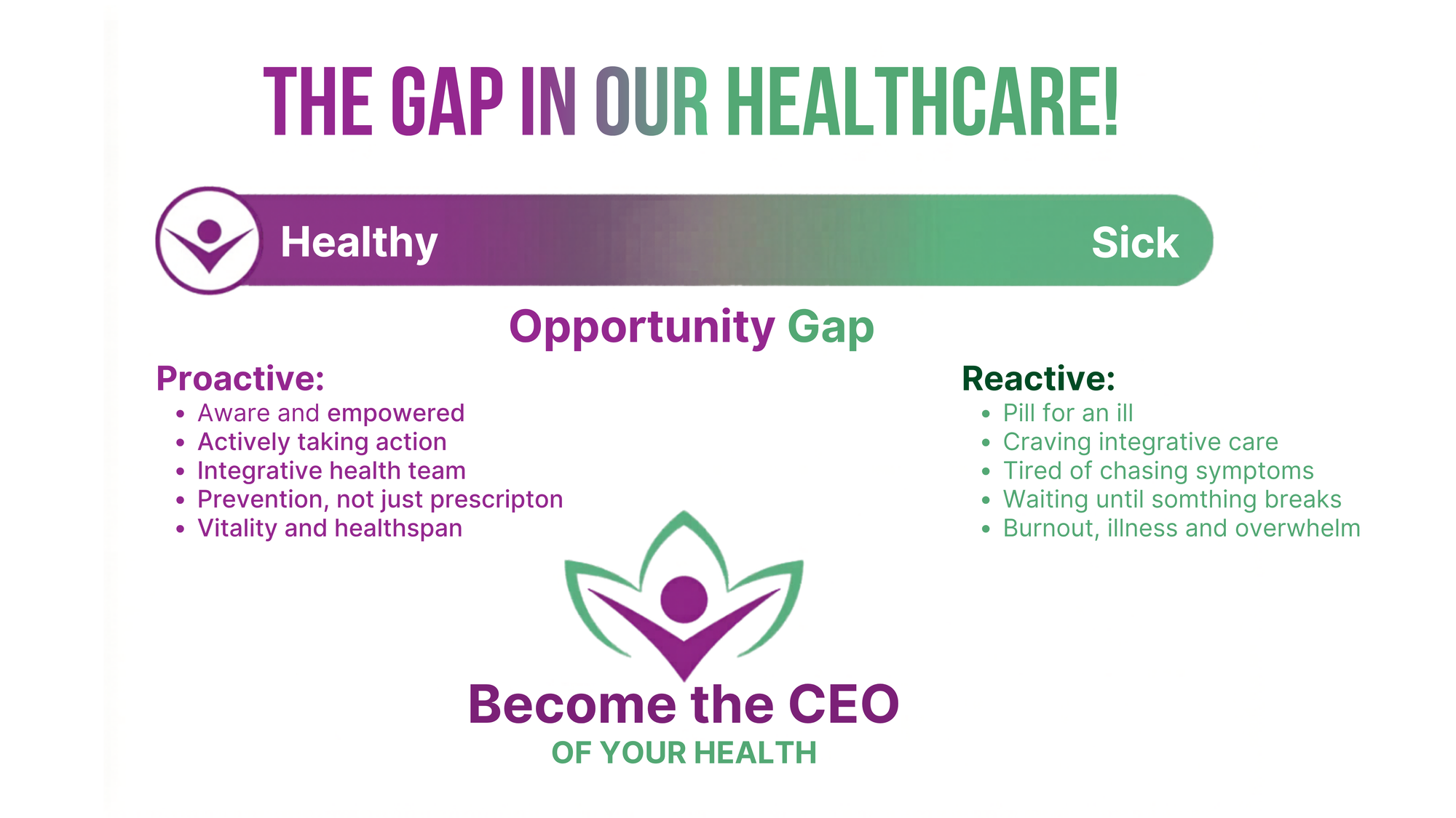
But there is still a long stretch between achieving peak bone mass and qualifying for routine publicly funded testing. I call it the opportunity gap: decades in which habits, hormones, health conditions and medications may be affecting bone but many people have no baseline, no feedback and no reason to believe they should be paying attention and getting into action.
And in Ontario, the opportunity gap widened in 2026
On April 1, 2026, OHIP introduced significant new restrictions on the frequency of publicly funded follow-up BMD testing.
This must be explained carefully. Ontario did not create a universal rule that everyone must wait until 70 for a first DXA. The age-70 baseline is the national clinical recommendation for people without risk factors, and an Ontario healthcare provider can still determine that an earlier baseline BMD is medically appropriate.
The sharper problem is what happens after that first test.
Under the new OHIP rules, a person considered high risk, defined for funding purposes as having a calculated 10-year fracture risk above 15%, can generally receive a repeat Bone Mineral Density (BMD) only once every 36 months. A repeat at 12 months is funded only for people with hypercortisolism or Cushing syndrome, or those receiving more than 20 mg per day of prednisone or its equivalent.
That narrow definition leaves important people caught in the middle. According to Osteoporosis Canada’s April 7, 2026 position statement, someone with a new fragility fracture, hyperparathyroidism, rheumatoid arthritis, a genetic metabolic bone condition, or rapid bone loss related to an aromatase inhibitor or androgen-deprivation therapy may still have to wait 36 months for another OHIP-funded DXA, even when the treating clinician believes earlier reassessment is warranted.
Osteoporosis Canada has stated plainly that the new OHIP access rules are not consistent with its 2023 clinical guideline and are not in the best interests of patient care. The organization’s concern is not that every person needs annual testing. It is that a rigid reimbursement rule can override clinical judgment and delay appropriate monitoring for people whose bone health may be changing quickly.
This is an essential distinction: a clinical guideline advises care; a funding schedule determines what the public system will pay for. They are not the same thing.
In my view, moving toward longer, one-size-fits-all testing intervals while bone loss remains silent is not a step toward prevention. It risks turning time, the very thing people need in order to act, into another barrier.
The false economy of waiting and the ageism we need to challenge
As a Certified Professional Consultant on Aging, I have long spoken up against ageism in healthcare for years. We must be willing to ask whether policies affecting older adults are being evaluated only through the cost of providing a test or through the far greater cost of what can happen when prevention and timely monitoring are missed.
The World Health Organization defines ageism as stereotypes, prejudice and discrimination based on age, and notes that it can be embedded within institutions as well as individual interactions. It is associated with poorer health, reduced recovery from disability, lower quality of life and earlier death. (World Health Organization)
I am not claiming that Ontario’s BMD policy was created because of ageism, nor has the government stated that the changes were made simply to save money. But when access is restricted largely within an aging population, we have a responsibility to ask hard questions: Are we treating longer life as a burden to manage or as added years worth protecting? Are we measuring the cost of prevention while ignoring the cost of waiting?
A fracture is not a single event on a balance sheet. It may mean ambulance and emergency care, surgery, hospitalization, rehabilitation, home care or admission to long-term care. It can also mean time away from work, lost productivity and unpaid caregiving by family members.
Canadian research using 2010–2011 health-system data estimated the annual burden of osteoporosis at more than $4.6 billion in 2014 dollars. That included 131,443 osteoporosis-attributable fractures, almost 65,000 acute-care admissions and more than 983,000 hospital days, as well as rehabilitation, home care and long-term care. This is not a current-dollar estimate, but it exposes the scale of the false economy: fractures are extraordinarily expensive. (Hopkins et al., 2016)
The human cost is even harder to calculate.
A 2026 review in JAMA reported a median one-year mortality rate of 22% after a hip fracture. It also found that only about 42% to 71% of patients regain their pre-fracture level of basic daily function within six months. That does not mean a hip fracture directly causes every death, and it does not mean every fracture carries the same risk. It does mean that hip fractures are serious, life-altering events associated with lost mobility, lost independence and reduced quality of life. (Johannesdottir et al., 2026)
Behind every statistic is a person who may no longer be able to live alone, return to work, travel, care for a partner or pick up a grandchild.
Prevention is not merely a healthcare expense. It is an investment in independence, participation, productivity and human dignity.
Prevention lives in that gap.
It is where we can ask earlier questions, identify modifiable risks and take informed action to support bone and muscle before a life-changing fracture forces the conversation.
Bone is not a dead frame
We often think of the skeleton as scaffolding: hard, silent and static. In reality, bone is living, metabolically active tissue that is continually being broken down and rebuilt.
Your skeleton does far more than give your body shape. It:
- protects the brain, spinal cord and vital organs;
- provides the levers and attachment points that allow muscles to create movement;
- stores and helps regulate minerals, including calcium and phosphorus;
- houses bone marrow, where blood cells are produced; and
- works with muscle, balance and the nervous system to support mobility, performance and independence.
This is why I do not see bone health as an isolated issue or as an “older woman’s problem.” It is part of the foundation of healthspan: the years in which we can work, lift, travel, dance, recover from illness, play with our grandchildren and remain independent.
Muscle belongs in this conversation too. Strong muscles load bone, support balance and help us recover when we stumble. Bone loss and sarcopenia, the age-related loss of muscle mass and function, can amplify one another, increasing the risk of falls, frailty and loss of independence. We need to stop discussing bone and muscle as if they live in separate bodies.
Performance is not reserved for elite athletes
When people hear the word performance, they often picture a podium, a finish line or a professional athlete. But performance belongs to all of us.
Performance is having the strength to climb the stairs, carry groceries and lift your grandchildren. It is being able to travel without your body dictating the itinerary. It is returning to the activities you love after illness or injury. It is continuing to work, garden, hike, golf, dance and move through the world with confidence and with as few restrictions as possible.
This is where bone health becomes a longevity conversation.
Longevity is not simply accumulating more birthdays. It is protecting the physical reserve that allows you to participate fully in those years. Healthy bone, adequate muscle, balance, mobility and the ability to recover all help determine whether a longer life is also an active and independent one.
Looking earlier gives us an opportunity to think beyond disease detection and toward health optimization. That does not mean chasing a “perfect” scan score or promising that every fracture can be prevented. It means understanding your baseline, recognizing risks, supporting what can be changed and monitoring your progress in the context of your health, goals and stage of life.
For an athlete, that conversation may be about training, recovery, under-fuelling or returning safely from injury. For everyone else, it may be about maintaining the capacity to live, work, travel and care for the people we love.
That is everyday performance. And it may be one of the most meaningful measures of healthspan we have.
What can place bone health at risk?
Age is only one part of the story. Bone can be affected by many factors across a lifetime, including:
- menopause, early menopause and other causes of low estrogen or testosterone;
- low body weight, under-fuelling and eating disorders;
- low physical activity or prolonged immobility;
- low calcium, vitamin D or protein intake;
- type 1 and type 2 diabetes, particularly long-standing type 2 diabetes, which can compromise bone quality and increase fracture risk even when bone density appears normal or higher;
- celiac disease, inflammatory bowel disease and other conditions that affect nutrient absorption;
- rheumatoid arthritis and other chronic inflammatory diseases;
- thyroid, parathyroid, kidney and liver disorders;
- long-term systemic glucocorticoid use, such as prednisone;
- some cancer treatments, including aromatase inhibitors for breast cancer and androgen-deprivation therapy for prostate cancer;
- some antiseizure medications; and
- smoking and higher alcohol intake.
Proton-pump inhibitors have also been associated with fracture risk in observational research, particularly with long-term or high-dose use, but association is not the same as proof of cause.
No one should stop a prescribed medication because of a scan or a blog. Medication risks and benefits belong in a conversation with the prescribing clinician.
The important point is this: bone health has a history. Your scan result should never be interpreted without your medical history, medications, nutrition, movement, hormones, previous fractures and goals.
A different way to look: what REMS adds
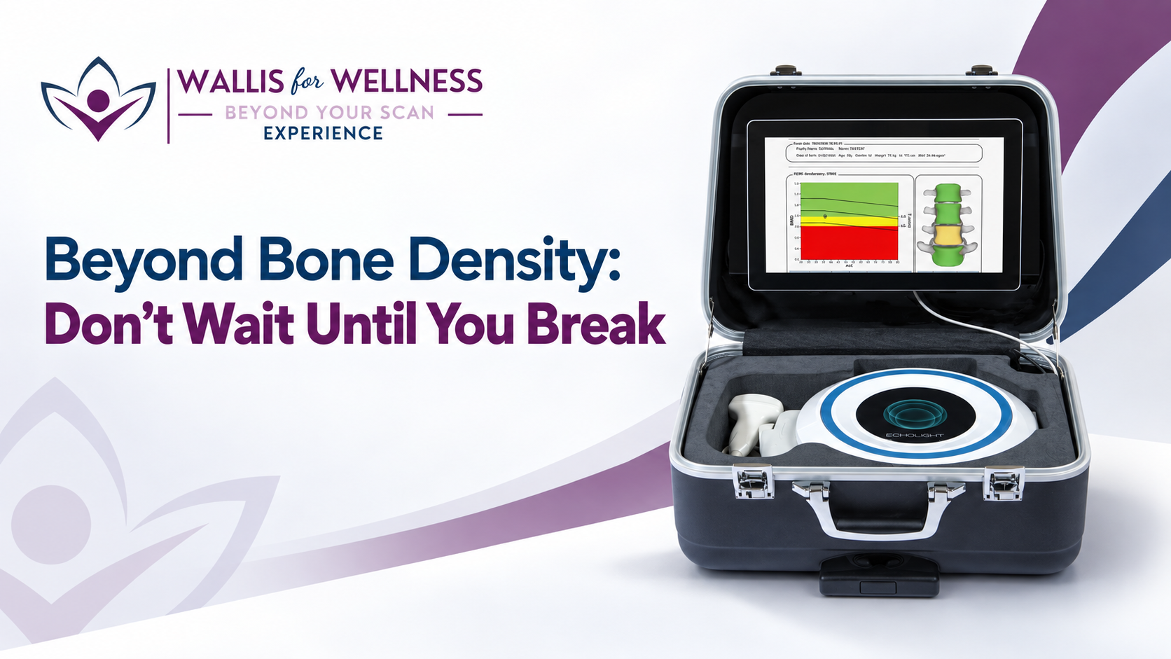
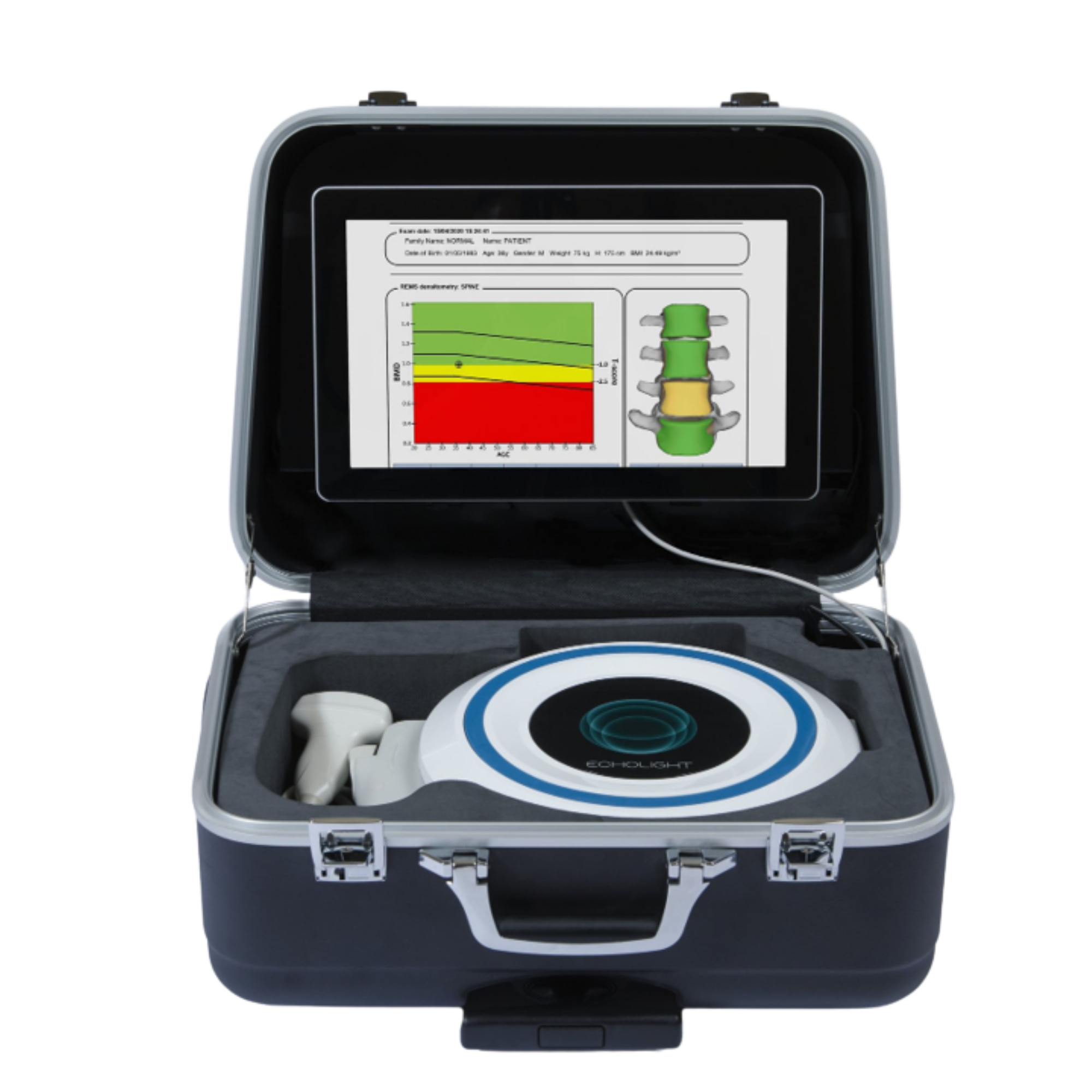
Echolight REMS stands for Radiofrequency Echographic Multi Spectrometry. It is a non-ionizing ultrasound-based technology that assesses the lumbar spine and proximal femur (hip) the same central skeletal sites commonly assessed by DXA.
Instead of using X-rays, REMS analyzes the raw radiofrequency signals returned from the bone during an ultrasound scan. Its software compares the spectral characteristics of those signals with reference models to estimate bone mineral density and generate diagnostic parameters.
Bone density and bone quality are not the same thing
For decades, DXA was essentially the only game in town. It gave us a valuable measure of bone mineral density: how much mineral is contained within a measured area of bone. But the amount of bone is only one part of what determines whether that bone can withstand force.
Bone quality is the broader story of how the bone is built and behaves. It includes microarchitecture, the internal arrangement of the bone, as well as turnover, mineralization and accumulated microscopic damage. Density and quality are related, but they are not interchangeable. Both contribute to bone strength and fracture risk.
Because I am a visual learner, I often compare it to a brick wall and the Eiffel Tower.
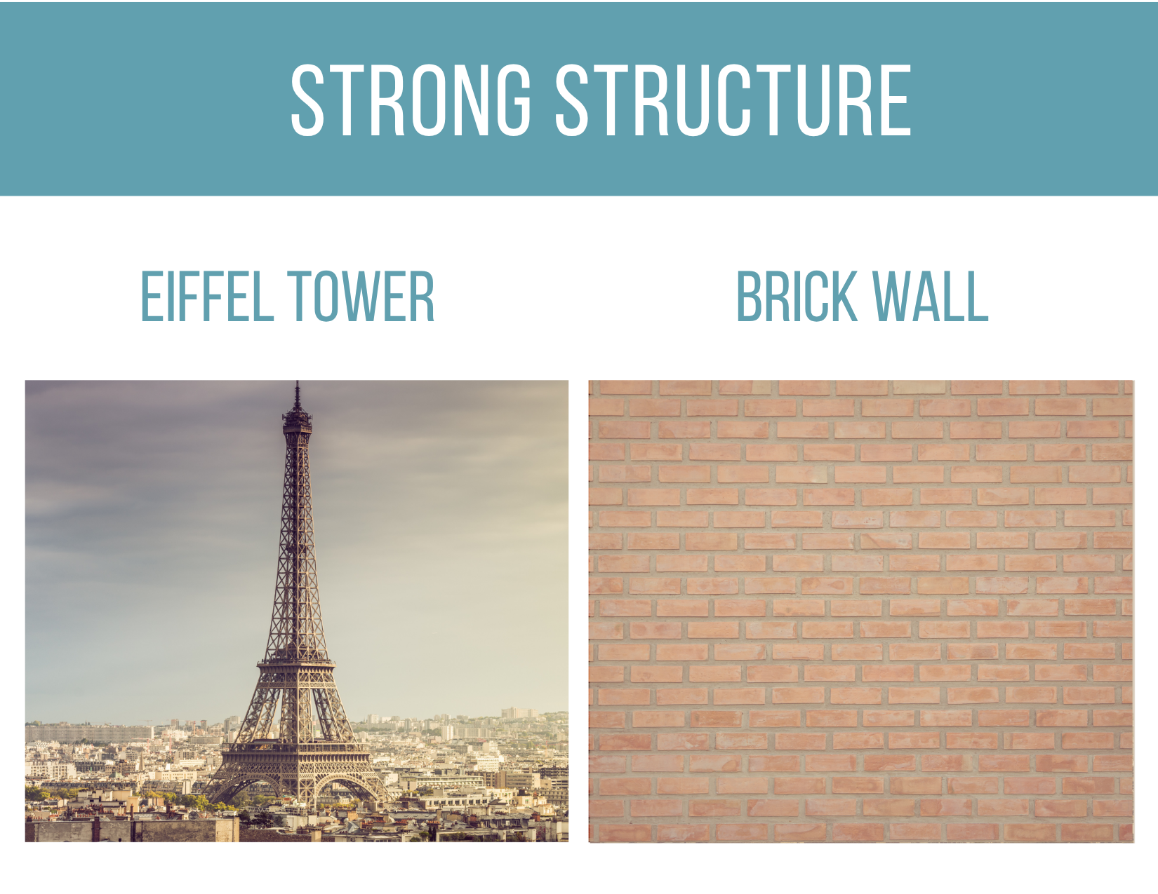
A brick wall can look thick, solid and dense. But if the mortar is deteriorating and the foundation is crumbling, its apparent mass can be deceiving. The Eiffel Tower uses far less material and contains open space, yet the quality, organization and engineering of its steel framework allow it to carry enormous forces.
Bones are not buildings, and this analogy is not a diagnostic model. But it helps illustrate an important point: knowing how much material is present does not, by itself, tell us everything about how well the structure may resist stress. A fuller assessment considers density alongside the characteristics associated with fragility, the person’s clinical risk factors and their health history.
This is where REMS offers an additional perspective. Its Fragility Score does not directly photograph or measure bone microarchitecture. Instead, it analyzes characteristics within the ultrasound signals that research has associated with fragile or more resilient bone patterns. It is one more piece of information, not a stand-alone verdict, to place alongside bone density and the person’s complete clinical picture.
REMS is:
- radiation-free;
- portable;
- designed to assess central skeletal sites; and
- able to provide an additional ultrasound-based Fragility Score, intended to capture aspects of bone fragility beyond bone mineral density alone.
Pregnancy, implants and degenerative changes: what REMS can and cannot assess
The absence of ionizing radiation creates opportunities for people who may not be suitable for, or well served by, conventional imaging but the details matter.
Pregnancy is one of those opportunities. Echolight’s current manufacturer guidance states that REMS is considered safe during pregnancy because it uses ultrasound rather than ionizing radiation. During pregnancy, however, the manufacturer specifies that the examination is performed at the femoral site only, keeping the scan area away from the abdomen and fetus. This makes REMS a potential option for evaluating maternal bone health when clinically appropriate, but it should never be promoted as an unrestricted full spine-and-hip examination during pregnancy. (Echolight FAQ)
Pregnancy and especially breastfeeding, temporarily changes how the body uses calcium and remodels bone to support the baby. Most women rebuild this bone after weaning, but some enter pregnancy with lower reserves or additional risk factors, and rare cases of pregnancy and lactation-associated osteoporosis can result in fractures. When clinically appropriate, radiation-free REMS gives women an opportunity to look sooner during pregnancy using the specified femoral-only protocol or after delivery, so they can better understand and support their long-term bone health. Pregnancy and lactation can uncover an existing vulnerability that otherwise might remain invisible. (Kovacs, 2025; Gak et al., 2024)
REMS may also be useful when degenerative changes or certain types of orthopaedic hardware can complicate DXA interpretation. Echolight explains that its software can identify ultrasound signals associated with artifacts, including osteophytes and exclude the affected portion from the BMD calculation.
Published REMS practice parameters also describe the exclusion of signals associated with calcifications, implanted metalwork and orthopaedic cement. This may help reduce the falsely elevated BMD readings that degenerative changes can sometimes produce with DXA. It does not mean that every degenerative or sclerotic condition can automatically be scanned or interpreted without limitation.
Hardware must be considered site by site. REMS may be able to assess a lumbar spine containing rods, screws or vertebral cement, or a hip containing certain fracture-fixation hardware, if enough usable bone tissue remains for a valid analysis. A total or partial hip replacement itself cannot be assessed. When one hip has been replaced, the opposite, unreplaced hip and the lumbar spine may still be evaluated when technically and clinically appropriate. (Zambito et al., 2025)
This is precisely why innovation must be paired with trained acquisition by a sonographer, careful screening and qualified interpretation. The right question is not simply, “Can REMS scan this person?” It is, “Which anatomical site can be assessed reliably, under the manufacturer’s protocol, and what can the resulting data legitimately tell us?”
That last point matters because bone strength is not determined by density alone. Bone geometry, microarchitecture, turnover, mineralization and accumulated damage all contribute to how well bone resists fracture. A person can sustain a fragility fracture without having a DXA T-score in the osteoporosis range of -2.5 standard deviations from normal healthy 30 yr old bone or greater.
Research comparing REMS with DXA has reported promising diagnostic agreement and precision across diverse populations. A large multicentre study found high correlation between REMS and DXA measurements and sensitivity and specificity above 90% at the lumbar spine and femoral neck.
A later European multicentre study also reported good diagnostic performance across five countries. A five-year prospective study found that REMS measurements predicted incident fragility fractures and, in that study population, identified fracture risk at least comparably to DXA. (Di Paola et al., 2019; Cortet et al., 2021; Adami et al., 2020)
More recent expert reviews describe REMS as a promising, portable and radiation-free option for bone assessment while also emphasizing the need for continued independent research, wider clinical experience and clear integration into established care pathways. (Fuggle et al., 2024)
That is the responsible way to talk about innovation: neither dismiss it because it is newer, nor crown it as magic because it is exciting.
REMS does not replace clinical judgment. It does not diagnose every cause of poor bone health. It is not a crystal ball, and it should not be positioned as a universal replacement for DXA. It is another evidence-informed way to look, one that can help open an earlier, richer conversation about bone strength and fracture prevention.
Why “Beyond Your Scan” Experience had to exist
Over the past several years, I have had the privilege of providing post-scan education to thousands of people. Again and again, I saw the same problem.
People received numbers, graphs and labels but were all left asking:
What does this mean for me? What do I do now? Who should I speak with? What questions should I ask?
A report without understanding can create fear. A report placed in context can create agency.
That is why Beyond Your Scan is not simply an appointment with a device. It is an experience built around four steps:
- Look. Your REMS assessment is performed by a trained professional, whether you visit our headquarters or attend a community, clinic or corporate location near you. Because the technology is portable, we can bring this opportunity beyond the walls of a traditional imaging centre and into more of the places where people live and work.
- Understand. Immediately after your scan, you receive an educational consultation to help you understand the reports in plain language what the findings mean, what they do not mean and which questions deserve further discussion. The goal is to help you articulate your results more confidently when speaking with your physician and integrative healthcare team.
- Learn and advocate. You receive private access to a multimedia-rich educational portal filled with videos, research, practical resources and a self-care toolbox to support your musculoskeletal and overall health. This is where we begin answering the question that matters most: “Now what?” Knowledge gives you better questions, and better questions help you become a more informed advocate for your health.
- Connect and build your team. We help you consider which types of qualified healthcare professionals may belong on your personalized team and, where appropriate, provide options you can explore.
The Preventative Health Awareness Movement (PHAM), our growing PHAMily of more than 100+ diverse healthcare professionals across North America, is united by a belief in prevention, collaboration and patient empowerment. No single practitioner holds every answer. The power lies in helping you assemble the right team for your needs, goals and health story.
This is not “scan, report and goodbye.” It is technology wrapped in education, resources, self-advocacy and human connection, so you are better equipped to help yourself and Become the CEO of your Health.
Clinical leadership grounded in experience, prevention and collaboration & medical insight.
Wallis for Wellness: Beyond Your Scan is honoured to have Dr. Kristy Prouse, BA, BSc, MD, FRCSC, as our Chief Medical Advisor.
Beyond Your Scan is a vision I have built from more than 34 years of experience as a Registered Massage Therapist, healthcare educator and advocate for preventative, integrative care.
As a regulated health professional, the former owner of an award winning multidisciplinary health clinic and the founder of the Preventative Health Awareness Movement, I have spent my career helping people better understand their bodies, ask more informed questions and connect with the appropriate healthcare professionals. I have always believed that people deserve to participate meaningfully in decisions about their health.
That being said, I have always stayed in my lane and respected my scope of practice. We do not prescribe where we are not authorized to prescribe, and we do not pretend one practitioner has every answer.
That is not fragmentation. That is responsible collaboration. That is effective patient centred healthcare.
I am honoured to have Dr. Kristy Prouse, BA, BSc, MD, FRCSC, serving as Chief Medical Advisor to Wallis for Wellness: Beyond Your Scan.
Dr. Prouse is an obstetrician-gynaecologist and hormonal-health physician whose career reflects a shared commitment to prevention, patient education and collaboration across conventional, functional and integrative healthcare. She was willing to think differently before these approaches became part of the wider healthcare conversation, and she understands that innovation must always be paired with medical responsibility.
Together, Dr. Prouse and I bring complementary perspectives: medical insight, decades of frontline clinical experience, patient education and a shared belief in collaborative care.
We are not here to prescribe someone’s path or create their treatment plan. We provide understandable information, educational resources and access to qualified professionals so each person can ask better questions, work with their own healthcare team and make informed decisions about what comes next.
We provide the knowledge, tools and connections. You remain at the centre of your care and in the driver’s seat of your health.
Being the CEO of your Health does not mean diagnosing yourself, rejecting conventional medicine or trying to manage everything alone. A good CEO does not do every job in the organization. A good CEO asks better questions, gathers the right team, understands the available information and participates in informed decisions.
That is health esteem: not believing you can control every outcome, but believing your health is worth understanding, protecting and advocating for.
Do not wait until you break
The future of healthcare will not be built by one device, one profession or one philosophy of care. It will be built by connecting good technology with sound clinical judgment, meaningful education and genuine collaboration.
Your bone health is part of your whole health. It changes across a lifetime. It responds to movement, nutrition, hormones, illness, medication and time. And because it is largely silent, it deserves our attention before it demands it.
The scan gives us a place to begin.
Beyond Your Scan Experience helps you decide what comes next.
We cannot wait for you to experience Wallis for Wellness: Beyond Your Scan.
Look sooner. Learn more. Build your team. And step into the driver’s seat of your health.
Book your REMS bone health assessment today. Don’t wait until you break.
Book your scan 🦴💪
A note about your care
This article is for education only and is not medical advice. REMS results should be interpreted in the context of your medical history and discussed with an appropriately qualified healthcare professional. Do not begin, stop or change medications or treatment based solely on a REMS result. Eligibility, contraindications and the appropriate timing of repeat assessment should be determined according to the device’s authorized use and your individual clinical circumstances.
References
- Morin SN, Feldman S, Funnell L, et al. Clinical practice guideline for management of osteoporosis and fracture prevention in Canada: 2023 update. CMAJ. 2023. Osteoporosis Canada guideline and tools.
- Di Paola M, Gatti D, Viapiana O, et al. Radiofrequency echographic multispectrometry compared with dual X-ray absorptiometry for osteoporosis diagnosis on lumbar spine and femoral neck. Osteoporosis International. 2019;30:391–402. DOI.
- Cortet B, Dennison E, Diez-Perez A, et al. Radiofrequency Echographic Multi Spectrometry (REMS) for the diagnosis of osteoporosis in a European multicenter clinical context. Bone. 2021;143:115786. PubMed.
- Adami G, Arioli G, Bianchi G, et al. Radiofrequency echographic multi spectrometry for the prediction of incident fragility fractures: a 5-year follow-up study. Bone. 2020;134:115297. DOI.
- Fuggle NR, Curtis EM, Ward KA, et al. Radiofrequency echographic multi spectrometry (REMS) in the diagnosis and management of osteoporosis: state of the art. Osteoporosis International. 2024. PubMed.
- National Institute of Arthritis and Musculoskeletal and Skin Diseases. Osteoporosis: causes, risk factors and bone biology. NIAMS.
- Osteoporosis Canada Scientific Advisory Council. Changes in BMD Testing in Ontario. April 7, 2026. Position statement.
- GraceMed. Dr. Kristy Prouse: Hormonal Health and OB/GYN Expert. Professional biography.
- Hopkins RB, Burke N, von Keyserlingk C, et al. The current economic burden of illness of osteoporosis in Canada. Osteoporosis International. 2016;27:3023–3032. DOI.
- Johannesdottir F, Roberts JE, Kiel DP, Tsai JN. Hip Fractures: A Review. JAMA. 2026;336(6):496–507. PubMed.
- World Health Organization. Ageism: overview, health effects and global response. WHO.
- Echolight. Frequently Asked Questions: pregnancy safety and osteophyte handling with REMS. Echolight FAQ.
- Zambito K, Kushchayeva Y, Bush A, et al. Proposed practice parameters for the performance of radiofrequency echographic multispectrometry (REMS) evaluations. Bone & Joint Open. 2025;6(3):291–297. DOI.